Urethrogram
What are the generally accepted indications for a urethrogram? A urethrogram is a procedure used to show the location and…
Read more


Radiography is the imaging of body structures, or parts of the body, using X-rays. X-rays are a form of radiation (X-radiation) similar to visible light, radio waves and microwaves. X-radiation is special because it has a very high energy level that allows the X-ray beam to penetrate through the body and create an image or picture.
Plain X-rays are the simplest medical images created through X-radiation. Other tests that also use X-rays are more complicated and require the use of computers to generate a picture; for example, computed tomography: CT (see InsideRadiology: CT scan). Any image created using an X-ray is due to different X-radiation absorption by different structures or parts in the body. A dense structure, such as bone, absorbs a high percentage of the X-ray beam (which appears light grey on the image), whereas low-density structures, such as soft tissues, absorb a small percentage (which appears dark grey on the image). The body has many different structures of varying densities and this difference creates a picture or image (see Figure 1).
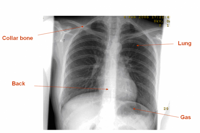
Figure 1: Chest X-ray. Bone (collar bone and back) appear light grey. Air (lungs and stomach gas) appears dark grey.
An X-ray examination is simple, quick and readily available at all radiology facilities. Your doctor might refer you for an X-ray if it is believed that an image of a certain part of your body could help in finding the cause of your symptoms and helping treatment. Most common X-rays are pictures of the chest (looking at the heart and lungs) and pictures of the arms, legs or spine in patients who have symptoms in the bones, joints or back.
No specific preparation is required for a plain X-ray.
It is important that you tell your own doctor and staff at the radiology facility where you are having the X-ray if there is any chance you might be pregnant. This is important information, as it will make a difference in the way the X-ray is carried out or a different test altogether might be required. Your safety and that of your unborn child is the number one priority.
You will usually be given a hospital gown to wear, as some clothing can make it difficult to see the images clearly. You might also need to remove certain items, such as watches, necklaces and some types of clothing that contain metal objects, such as zips.
If you are attending a follow-up X-ray to assess the progress of an injury or illness, you might need to take any previous X-rays with you so the radiologist (specialist doctor) who will send a report to your own doctor can compare the new X-ray with the old one and see if there have been any recent changes.
The radiographer (trained X-ray technologist) who will carry out the X-ray will explain the procedure to you. Depending on the part of your body being examined, your position (e.g. standing, sitting or lying), the number of X-rays taken and the speed of the test will vary.
It is important that you stay completely still when the radiographer instructs you to, as any movement might create a blurred image.
After the X-rays are completed, the radiographer will electronically process each X-ray and check the results for quality. While this is being done, which does not take very long, you will be asked to wait in the X-ray room or change room in your hospital gown. Sometimes there will be a need for additional images to be taken to obtain more information to help the radiologist (specialist doctor) to make a diagnosis. There is no need for concern if this happens, as it is quite common. In most cases, the extra X-rays are carried out to obtain a better view of the part of your body being examined, not because there is a problem.
The radiographer will instruct you when the procedure is finished. You may wish to ask them when the results will be available.
The radiologist then carefully assesses the images, makes a diagnosis and produces a written report on the findings. This report is sent to your doctor, specialist or allied health professional who referred you for the test.
You are welcome to ask questions about the process at any stage if you have any concerns.
The entire process is straightforward, and you will not feel anything strange or feel any different during the examination.
If you are bringing a child for an X-ray, you might be asked to help in settling or keeping the child in the correct position for the test. If you are asked to remain in the X-ray room, the radiographer will take all possible steps to ensure that you are not exposed to any X-rays.
You will not feel anything while the X-ray is being taken or afterwards.
It usually takes less than 15 minutes for an entire X-ray procedure. This depends on the number of parts of your body being examined and your mobility; that is, your ability to move about, and your general health. In most cases, the area being examined needs to be viewed from different directions to obtain enough information to make the diagnosis, and this might require you to move into different positions.
For example, a simple chest X-ray on an able and willing patient could take less than 1 minute. A distressed patient needing X-rays of the whole spine, pelvis, both shoulders and both legs could take 45 minutes.
People with disabilities and children will also take longer, particularly if they find it difficult to keep still or to cooperate with or understand instructions given by the radiographer carrying out the X-ray examination.
Generally, the benefit of the X-ray procedure is far more important than the small estimated risk of the effects of radiation. At the radiation dose levels that are used in diagnostic radiography, there is little or no evidence of health effects (for further information see InsideRadiology: Radiation risk of medical imaging for adults and children).
The type of radiation used in X-rays is called ionising radiation. Medical research has been unable to establish conclusively that there are significant effects for patients exposed to ionising radiation at the doses used in diagnostic X-ray imaging.
Radiographers are trained to use the smallest possible amount of X-rays required to produce a satisfactory image.
X-ray imaging is useful to diagnose disease and injury, such as pneumonia, heart failure, fractures, bone infections, arthritis, cancer, blockage of the bowel, collapsed lung and so on. It is fast and easy, so it is particularly useful in emergency diagnosis and treatment.
X-ray equipment is widely available in hospitals, X-ray clinics and other locations, making it convenient for both patients and doctors, even in remote locations.
A radiographer or medical imaging technologist is a trained health professional who carries out diagnostic radiography.
A radiologist is a specialist medical doctor who reviews and interprets the images and provides a written report of the test to your referring doctor, specialist or allied health worker.
Plain radiography/X-rays are carried out in the diagnostic imaging (radiology) department of most hospitals (although this depends on the size of hospital, as some small hospitals do not have X-ray facilities). They can also be carried out at special X-ray clinics, private radiology practices and other locations.
The time that it takes your doctor to receive a written report on the X-ray examination will vary, depending on:
You can ask the facility where you are having your X-ray when your doctor is likely to have the written report.
It is important that you discuss the results with the doctor or other health professional who referred you, either in person or on the telephone, so that they can explain what the results mean for you.
X-rays are safe when carried out in a controlled environment, such as an X-ray department. X-ray equipment is checked regularly to ensure that it is functioning properly and not delivering excess radiation to patients or staff. People operating X-ray equipment are required by law to be licensed to do so, to ensure they are properly qualified to operate the radiation equipment.
If you require any more information or have queries about your X-ray procedure, then please contact your local doctor or the X-ray facility where you have been referred for the X-ray examination.
Page last modified on 31/8/2017.
RANZCR® is not aware that any person intends to act or rely upon the opinions, advices or information contained in this publication or of the manner in which it might be possible to do so. It issues no invitation to any person to act or rely upon such opinions, advices or information or any of them and it accepts no responsibility for any of them.
RANZCR® intends by this statement to exclude liability for any such opinions, advices or information. The content of this publication is not intended as a substitute for medical advice. It is designed to support, not replace, the relationship that exists between a patient and his/her doctor. Some of the tests and procedures included in this publication may not be available at all radiology providers.
RANZCR® recommends that any specific questions regarding any procedure be discussed with a person's family doctor or medical specialist. Whilst every effort is made to ensure the accuracy of the information contained in this publication, RANZCR®, its Board, officers and employees assume no responsibility for its content, use, or interpretation. Each person should rely on their own inquires before making decisions that touch their own interests.